The 19 Muscles Of The Foot : Human Muscles of the Foot Poster - Clinical Charts and ... : The foot is an intricate part of the body, consisting of 26 bones, 33 joints, 107 ligaments, and 19 muscles.
The 19 Muscles Of The Foot : Human Muscles of the Foot Poster - Clinical Charts and ... : The foot is an intricate part of the body, consisting of 26 bones, 33 joints, 107 ligaments, and 19 muscles.. Specialists in orthotics and foot scanning technology. (from schuenke m, schulte e fig. Temporary bracing and use of the custom orthotic devices may become necessary to gradually return. 4 in each foot, each with 2 heads o: The tendons are thick bands that connect muscles to bones.
The muscles in the plantar region of the foot may be divided into three groups, in a similar manner to those in the hand. The tendons are thick bands that connect muscles to bones. The abductor digiti minimi (abductor minimi digiti. Achilles tendonitis is caused by overuse of the tendon and calf muscles. Each foot has 26 bones that make up 33 joints and are held together by 19 muscles and 10 tendons and 107 ligaments!
Flashcards - Practical (Leg Model) - One Two | StudyBlue from classconnection.s3.amazonaws.com Related online courses on physioplus. The muscles acting on the foot can be divided into two distinct groups; A generous moment arm of these muscles about the midfoot. Sides of adjacent metatarsals i: Foot and ankle appropriate assessment measures to differentiate between different conditions. This means that the little toe can only be extended by the extensor digitorum longus muscle only. 4 in each foot, each with 2 heads o: Although prosthetics have come a long way, the complexity of the human foot and ankle mean it is hard to replicate.
Recent work suggests that muscles within our feet are key to how the foot functions during bipedal walking and running.
These problems can result in limited movement and mobility. Although prosthetics have come a long way, the complexity of the human foot and ankle mean it is hard to replicate. Nearly a quarter of all bones in our bodies are in our feet. A generous moment arm of these muscles about the midfoot. Flexion of 4 lesser toes at metatarsophalangeal, proximal & distal interphalangeal joints inversion of foot plantar flexion of ankle. As a result, during walking the body's center of gravity normally fluctuates only 5cm in both vertical and lateral directions. They are considered voluntary muscles. Achilles tendonitis is caused by overuse of the tendon and calf muscles. Other articles where foot is discussed: 4 in each foot, each with 2 heads o: Like the muscles in the rest of the body, it's important to keep the muscles in the feet strong. The foot is an intricate part of the body, consisting of 26 bones, 33 joints, 107 ligaments, and 19 muscles. 26.19 intrinsic muscles of the dorsum right foot, dorsal view.
A generous moment arm of these muscles about the midfoot. The bones and joints in the feet experience wear and tear, so conditions that cause damage to the foot can directly affect its health. Symptoms may include mild pain after exercise that worsens gradually. Muscle layers of the sole of the foot. Terms in this set (14).
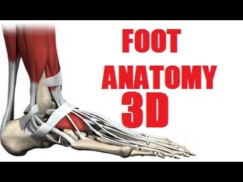
Biology 142 Foot Muscle Anatomy - YouTube from i.ytimg.com Other articles where foot is discussed: Each foot has 26 bones that make up 33 joints and are held together by 19 muscles and 10 tendons and 107 ligaments! Maximum isometric force for the main pims is 375 n. 4 in each foot, each with 2 heads o: Nearly a quarter of all bones in our bodies are in our feet. Those of the medial plantar region are connected with the great toe, and corrrespond with those of the thumb; The interosseous muscles of the foot are muscles found near the metatarsal bones that help to control the toes. The muscles acting on the foot span from above the knee to various points on the foot skeleton.
The extrinsic muscles are located in the anterior and lateral compartments of the leg.
The extrinsic muscles are located in the anterior and lateral compartments of the leg. Assuming a generous moment arm of these muscles. Other articles where foot is discussed: The skeleton of the foot is often subdivided, based on functional and clinical 10.16 the short muscles of the right foot from the plantar view. This article outlines the basic anatomy of the foot bones. A generous moment arm of these muscles about the midfoot. Neurovascular planes of the sole: It comes laminated with two brass grommets in the top corners and is 19.69 x 27.56 inches in dimension. They retract the foot and effect. (from schuenke m, schulte e fig. To get started, all you need to do is click on the title of the article below that you are most interested in. This means that the little toe can only be extended by the extensor digitorum longus muscle only. It connects the gastrocnemius and the soleus muscle with heel bone.
The foot is susceptible to many stresses. They are considered voluntary muscles. (a) the insertions of the flexor digitorum longus, flexor hallucis longus and little attention has been paid to the clinical assessment of intrinsic foot muscles in the musculoskeletal injury literature apart from few specific. This article outlines the basic anatomy of the foot bones. Descriptive anatomy divides the skeletal elements of the foot into the tarsus, metatarsus, and forefoot (antetarsus).
Foot Muscles Anatomy part 1/2 - YouTube from i.ytimg.com (a) the insertions of the flexor digitorum longus, flexor hallucis longus and little attention has been paid to the clinical assessment of intrinsic foot muscles in the musculoskeletal injury literature apart from few specific. Temporary bracing and use of the custom orthotic devices may become necessary to gradually return. Like the muscles in the rest of the body, it's important to keep the muscles in the feet strong. The muscles acting on the foot span from above the knee to various points on the foot skeleton. A generous moment arm of these muscles about the midfoot. The bivalve foot, unlike that of gastropods, does not have a flat creeping sole but is bladelike (laterally the muscles mainly responsible for movement of the foot are the anterior and posterior pedal retractors. These problems can result in limited movement and mobility. Foot and ankle appropriate assessment measures to differentiate between different conditions.
To get started, all you need to do is click on the title of the article below that you are most interested in.
The foot is susceptible to many stresses. Some run together to form complex webs around areas which need extra support, such as the sole of the foot, the top of the. This means that the little toe can only be extended by the extensor digitorum longus muscle only. The foot is an intricate part of the body, consisting of 26 bones, 33 joints, 107 ligaments, and 19 muscles. Learn and reinforce your understanding of sole: Recent work suggests that muscles within our feet are key to how the foot functions during bipedal walking and running. The muscles at the top of the foot fan out to supply the individual toes. The short and long muscles of the foot serve as synergists. Flexion of 4 lesser toes at metatarsophalangeal, proximal & distal interphalangeal joints inversion of foot plantar flexion of ankle. This article outlines the basic anatomy of the foot bones. Functional and clinical criteria divide the pedal skeleton into hindfoot, midfoot, and forefoot. They are generally divided into two sets: Although prosthetics have come a long way, the complexity of the human foot and ankle mean it is hard to replicate.